
 |
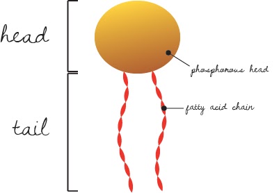
| Figure 1: A single phospholipid |
The membrane of a cell is made up of a phospholipid bilayer.
When you break those terms down, you can see it has something to do with
phosphorous, lipids, and two layers. Phospholipids are two-part molecules- on
one end, a phosphate group- called the head; on the other, two dangly chains of fatty acids- the tail (Fig. 1).
The phosphate head is polar, while the fat chains are
non-polar. Polar simply means that the charges within the molecules are more
clumped than evenly distributed. Polar molecules are attracted to other polar
molecules, like, oh say, water. As a result, the phosphate heads love to
snuggle up next to water, and always try to acclimate so that they are in
contact with it. As for those fat chains, well, they’re lipids. And we all know
that lipids (oils, fats, wax) repel water. Slap some fancy terminology in
there, and we have a molecule with one hydrophilic (water-loving) end and one
hydrophobic (water-fearing) end.
 |
| Figure 2: Phospholipid bilayer that forms a cell membrane |
The end result of all this is that phospholipids tend to form
sheets, where the hydrophobic fatty acids are sandwiched in between the
protective phosphate heads, which are exposed to the aqueous environments
inside and outside of the cell. These sheets form the shell of a sphere, and
are the cell membrane (Fig. 2).
Now, let’s apply this setup to
reality. The phospholipids are not cemented in place, and in fact shuffle
amongst each other like tightly-packed floating ducks in a bath tub. This
non-static membrane system is described by the fluid mosaic model. That
basically says that cell membranes are fluid (non-static) and mosaic in nature
(made up of sub-units). Ah lah, a membrane that is made up of subunits that
float and shift to form an ever-changing cell surface.
I like this cell membrane
business because it shows how a bunch of non-living molecules like phosphates
and fatty acids can work together to form a system that is living. Because of the fluid mosaic model and it’s important
duty of dividing non-life from life, the spectrum of “living” spans from an itty
bitty amoeba to a T-rex. As complex as we are as vertebrate organisms, it’s
important to remember that we are complexities made up of even more
complexities. Complexities like phospholipids, that are flashes of our humble
beginnings as single cells- a collection of working parts, housed together.
No comments:
Post a Comment